


The Impact of the Williams Syndrome Mutations on Neural James S. McDonnell Foundation Collaborative Activity Award: |
Project 4: Functional magnetic resonance imaging of social cognition in Williams syndrome individuals with full and small deletions (Bellugi and Raichle).
A critical component to the link between social behavior and genetics is an understanding of the functional brain activity of individuals with Williams syndrome. With the use of functional neuroimaging techniques that assess both basal metabolic activity as well as functional activity following social probes, this proposal seeks to further clarify the potential differences in neural activity in Williams individuals with large and small variants of the Williams deletion as compared with normal controls. Studies of human cognition have been at the forefront of brain imaging work for more than a decade and almost every aspect of human cognition has been examined. Studies related to social behavior would add a new dimension to the imaging agenda by bringing emerging imaging work on emotion into juxtaposition with studies of cognition. Work has amply shown the influence of limbic brain areas on behavior (Raichle, 1998). An important genetic dimension will be added by the study of the remarkable alterations in social behavior in Williams syndrome. Functional brain imaging will provide an important element of this overall program that includes studies of genetics, animal behavior and human disorders as they relate to human social behavior.
A series of experimental studies using social probes known to be relevant to Williams is planned. This component of the program focuses on the issue of whether the activity of certain brain regions may be associated with the hypersociability of the Williams syndrome subjects. We will use both fMRI indices of resting metabolism as well as stimulated activity with specifically designed probes of social cognition. The typical fMRI study measures the Blood Oxygenation Level Dependent (BOLD) effect which provides a sensitive tool for mapping patterns of activation in the human brain. However, the BOLD effect is not a pure reflection of changes in neuronal activity, nor even of changes in cerebral blood flow and has several potential drawbacks (Buxton, et al, 1997, 1998a,b). Thus we will include also a newly developed imaging technique based on arterial spin labeling that provides a direct measure of local cerebral blood flow. These studies will be carried out in collaborations between Dr. Raichle and the group at Washington University and the San Diego Brain Imaging group, including Richard Buxton. We also propose to assess functional brain activity in Williams syndrome with specific experimental paradigms for the neural underpinnings of social cognition.
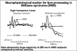 Recent work has shown that there is a distinctive neuroanatomical profile of the Williams brain that accompanies the unique cognitive and social phenotype (Reiss, et al, 2000; Galaburda & Bellugi, 2000). These findings of abnormalities in the volume and shape of the Williams brain, including the amygdala, suggest that there should be concomitant abnormalities in their functional activity as well. To begin to answer the question of how the Williams social cognitive phenotype with its hypersociability and affiliative behavior is represented in functional brain activity, we propose several fMRI experiments that will allow us to go across levels of analysis. Each of these studies will assess brain regions such as orbitofrontal cortex, fusiform gyrus and amygdala dysfunction in individuals with Williams. We now have data from ERP studies showing that these interesting alterations in social cognition do have a neural basis that can be detected by functional neuroimaging techniques and that some may differentiate Williams with different size deletions. As an example, Event Related Potential (ERP) studies have found that there is a neurophysiological marker for Williams in early brain wave responses to face processing, occurring in all full deletion Williams studied to date but not in normal controls or other groups (see Figure above). This marker may index the Williams increased attention to faces (Mills, et al, 2000) and provides a clue to differences in functional brain activity between Williams and normal controls in face processing, an important aspect of social behavior.
Significantly, a rare Williams individual with a small deletion who does not show the excessive sociability typical of Williams syndrome, also does not have the neurophysiological marker for face processing that is found with all other full deletion Williams studied to date (Korenberg et al, 2001; Korenberg et al, 2000). Instead, this subject’s brain activity during face processing is like that of normal controls. This finding provides a model for the brain activation studies proposed here, which will compare brain activation of typical and small deletion cases with normal controls.
Proposed fMRI experimental studies, contrasting groups of Williams with typical deletions, those with smaller deletions, and normal controls will center on particular dimensions of social cognition: an interest in faces, identification of faces and facial expression and a drive to approach strangers. One study builds on the experimental paradigm modified from Bellugi & Adolphs 1999, designed to assess the willingness with which individuals with bilateral amygdala damage approach a stranger for social interaction. Our behavioral studies have found that Williams individuals, like patients with bilateral amygdala damage, have higher social judgement scores than matched normal controls or individuals with brain damage in other regions. Therefore, we will assess the neural systems underlying social judgments such as the interest in approaching others and how these regions may potentially differ for individuals with varying Williams deletions and normal controls. Because the behavioral phenotype of Williams so distinctly involves the attraction to faces and a social interest in faces, understanding the role of such brain areas as the orbitofrontal cortex, amygdala, and fusiform gyrus in these aspects of social cognition may prove extremely fruitful.
In sum, the specific aim of the imaging studies in Project 4 is to determine the brain systems differentially altered in the typical and short forms of the Williams deletion and normal controls as clues to what regions might be involved in the hypersociability of Williams specifically and social cognition more generally. It is through the combination of functional imaging and molecular genetics, using Williams syndrome as a model, that we plan to "map" particular variations in brain activation for social cognition to specific genes, leading to the studies of gene expression in the brain.
| Introduction | Project 1 | Project 2 | Project 4 | Project 5 | Project 6 | Conclusion |